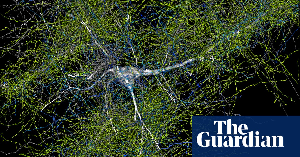
Scientists have reconstructed a wiring diagram for a piece of human brain in unprecedented detail, revealing fresh quirks and complexities in what many regard as the most sophisticated object in the known universe.
Harvard researchers teamed up with experts in machine learning at Google to map out the neural circuitry, connections, supporting cells and blood supply in a speck of healthy tissue removed from the cortex of a 45-year-old woman who had had surgery for epilepsy.
The clump of brain amounted to a mere cubic millimetre of tissue, but working out the wiring still presented a huge task for the team. Electron microscope images of more than 5,000 slices of the sample revealed 57,000 individual cells, 150m neural connections and 23cm of blood vessels.
“The aim was to get a high resolution view of this most mysterious piece of biology that each of us carries around on our shoulders,” said Jeff Lichtman, a professor of molecular and cellular biology at Harvard. “The reason we haven’t done it before is that it is damn challenging. It really was enormously hard to do this.”
Having sliced the tissue into wafers less than 1,000 times thinner than the width of a human hair, the researchers took electron microscope images of each to capture details of brain structure down to the nanoscale, or thousandths of a millimetre. A machine-learning algorithm then traced the paths of neurons and other cells through the individual sections, a painstaking process that would have taken humans years. The images comprised 1.4 petabytes of data, equivalent to 14,000 full length, 4k resolution movies.
“We found many things in this dataset that are not in the textbooks,” said Lichtman. “We don’t understand those things, but I can tell you they suggest there’s a chasm between what we already know and what we need to know.”
In one baffling observation, so-called pyramidal neurons, which have large branches called dendrites protruding from their bases, displayed a curious symmetry, with some facing forwards and others backwards. Other images revealed tight whorls of axons, the thin fibres that carry signals from one brain cell to another, as if they had become stuck on a roundabout before identifying the right exit and proceeding on their way.
The map also revealed rare instances where neurons made extremely strong connections with other cells. Across the whole lump of brain tissue, more than 96% of axons made only one connection with a target cell, with 3% making two connections. But a handful made tens of connections, and in one case more than 50, with a nearby cell. Details are published in the journal Science.
Lichtman speculated that such strong connections might help explain how well-learned behaviours – such as removing your foot from the accelerator and applying the brake at a red light – require almost zero thought after enough practice. “I think these powerful connections may be part of the system of learned information and what learning looks like in the brain,” he said. The team is making the map freely available for other researchers to use.
For now, the researchers are not even thinking about mapping a whole human brain. The task is too hard technologically, and healthy human brains do not grow on trees. Instead, the next project will be a multi-university collaboration with Google to reconstruct the wiring of an entire mouse brain. That might shed light on the brain circuits that make a mouse move towards Swiss cheese, and in turn what makes a human pause outside a bakery. “You would get some insight into how human will is guided by sensory experience,” said Lichtman. “There are really wonderful opportunities, if you have a whole mouse brain, to get insight into free will, even,” said Lichtman. “You know, a mouse is not a robot.”


