Blood is a remarkable material: it must remain fluid inside blood vessels, yet clot as quickly as possible outside them, to stop bleeding. The chemical cascade that makes this possible is well understood for vertebrate blood. But hemolymph, the equivalent of blood in insects, has a very different composition, being notably lacking in red blood cells, hemoglobin, and platelets, and having amoeba-like cells called hemocytes instead of white blood cells for immune defense.
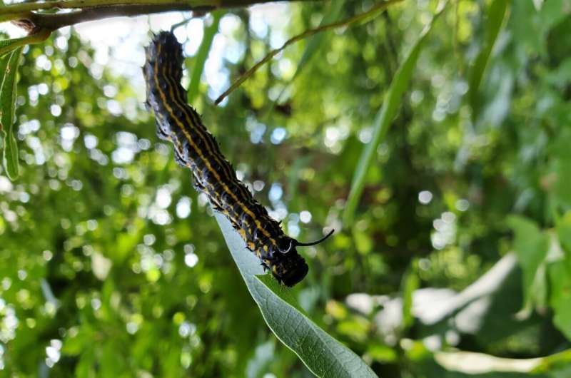
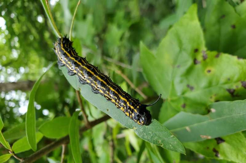
Just like blood, hemolymph clots quickly outside the body. How it does so has long remained an enigma. Now, materials scientists have shown in Frontiers in Soft Matter how this feat is managed by caterpillars of the Carolina sphinx moth. This discovery has potential applications for human medicine, the authors said.
“Here we show that these caterpillars, called tobacco hornworms, can seal the wounds in a minute. They do that in two steps: first, in a few seconds, their thin, water-like hemolymph becomes ‘viscoelastic’ or slimy, and the dripping hemolymph retracts back to the wound,” said senior author Dr. Konstantin Kornev, a professor at the Department of Materials Science and Engineering of Clemson University.
“Next, hemocytes aggregate, starting from the wound surface and moving up to embrace the coating hemolymph film that eventually becomes a crust sealing the wound.”
Challenging to study
Fully grown tobacco hornworms, ready to pupate, are between 7.5cm and 10cm long. They only contain a minute amount of hemolymph, which typically clots within seconds, which makes it hard to study with conventional methods.
For these reasons, Kornev and colleagues had to develop new techniques for the present study, and work fast. Even so, the failure rate for the trickiest manipulations was enormous (up to 95%), requiring many attempts.
They restrained individual hornworms in a plastic sleeve, and made a slight wound in one of each caterpillar’s pseudolegs through a window in the sleeve. They then touched the dripping hemolymph with a metal ball, which was pulled away, creating a hemolymph ‘bridge’ (about two millimeters long and hundreds micrometers wide) that subsequently narrowed and broke, producing satellite droplets. Kornev and team filmed these events with a high frame rate camera and macro lens, to study them in detail.

Instantaneous change in properties
These observations suggested that during the first approximately five seconds after starting to flow, hemolymph behaved similarly to water: in technical terms, like a Newtonian, low viscosity liquid. But within the next 10 seconds, the hemolymph underwent a marked change: it now did not break instantaneously but formed a long bridge behind the falling drop. Typically, bleeding stopped completely after 60 to 90 seconds, after a crust formed over the wound.
Kornev and colleagues studied the hemolymph’s flow properties further by placing a 10-micrometer-long nickel nanorod in a droplet of fresh hemolymph. When a rotating magnetic field caused the nanorod to spin, its lag relative to the magnetism gave an estimate of the hemolymph’s ability to hold the rod back through viscosity.
They concluded that within seconds after leaving the body, caterpillar hemolymph changes from a low-viscous into a viscoelastic fluid.
“A good example of a viscoelastic fluid is saliva,” said Kornev. “When you smear a drop between your fingers, it behaves like water: materials scientists will say it is purely viscous. But thanks to very large molecules called mucins in it, saliva forms a bridge when you move your fingers apart. Therefore, it’s properly called viscoelastic: viscous when you shear it and elastic when you stretch it.”
The scientists further used optical phase-contrast and polarized microscopy, X-ray imaging, and materials science modeling to study the cellular processes by which hemocytes aggregate to form a crust over a wound. They did this not only in Carolina sphinx moths and their caterpillars, but also in 18 other insect species.
Hemocytes are key
The results showed that hemolymph of all species studied reacted similarly to shear. But its reaction to stretching differed drastically between the hemocyte-rich hemolymph of caterpillars and cockroaches on the one hand, and the hemocyte-poor hemolymph of adult butterflies and moths on the other: droplets stretched out to form bridges for the first two, but immediately broke for the latter.
“Turning hemolymph into a viscoelastic fluid appears to help caterpillars and cockroaches to stop any bleeding, by retracting dripping droplets back to the wound in a few seconds,” said Kornev.
“We conclude that their hemolymph has an extraordinary ability to instantaneously change its material properties. Unlike silk-producing insects and spiders, which have a special organ for making fibers, these insects can make hemolymph filaments at any location upon wounding.”
The scientists concluded that hemocytes play a key role in all these processes. But why caterpillars and cockroaches need more hemocytes than adult butterflies and moths is still unknown.
“Our discoveries open the door for designing fast-working thickeners of human blood. We needn’t necessarily copy the exact biochemistry, but should focus on designing drugs that could turn blood into a viscoelastic material that stops bleeding. We hope that our findings will help to accomplish this task in the near future,” said Kornev.
More information:
To seal a wound, caterpillars transform blood from a viscous to a viscoelastic fluid in a few seconds, Frontiers in Soft Matter (2024). DOI: 10.3389/frsfm.2024.1341129. www.frontiersin.org/articles/1 … fm.2024.1341129/full
Citation:
Scientists discover how caterpillars can stop their bleeding in seconds (2024, March 27)
retrieved 27 March 2024
from https://phys.org/news/2024-03-scientists-caterpillars-seconds.html
This document is subject to copyright. Apart from any fair dealing for the purpose of private study or research, no
part may be reproduced without the written permission. The content is provided for information purposes only.


