Research groups led by Prof. Bi Guoqiang, University of Science and Technology of China (USTC), and Prof. Zhou Pengcheng from Shenzhen Institutes of Advanced Technology of Chinese Academy of Chinese proposed a design for ultra-compact head-mounted fluorescence microscopes, which were applied to neuro observations. The study was published in National Science Review.
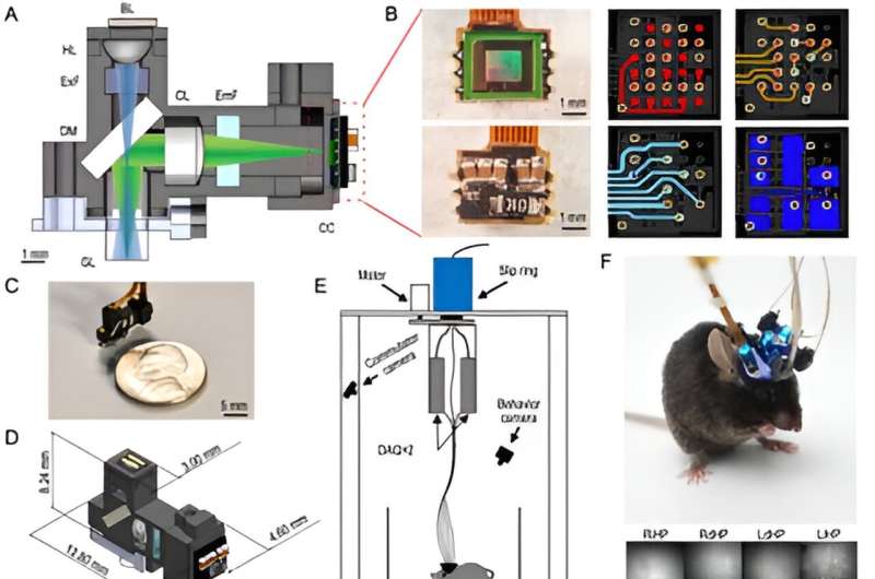
Head-mounted microscopes are applied for calcium imaging in freely behaving animals. However, space and weight constraints remain a barrier. In this study, researchers proposed a Tightly Integrated Neuronal Imaging microscope (TINIscope) with optimized optical, electronic and mechanic designs. Weighing 0.43g, TINIscope is the lightest among current head-mounted microscopes. Due to its compactness, the TINIscope is capable of multi-region calcium imaging.
Instead of using a serializer chip, the image sensor directly transmits serialized data, thus cutting much of the size. Some other components were also cut, with only essential parts left. The shrunk sensor allowed an optimized illumination design to reduce the distance between optical components further.
TINIscope applied a novel design with the placement of the CMOS sensor on the side and LED excitation on the top. With dichroic mirrors applied to reflect emission fluorescence to sensors, multiple TINIscopes could be rotated to distribute their sensors spatially. Therefore, multi-region imaging was reached. Finally, a commutator was developed to prevent entanglement caused by the motion of the mouse.
In the experiment, the calcium-sensitive fluorescent protein GCaMP6s was applied, and each of the four hippocampal subregions of a mouse was implanted with a GRIN lens. The equipment of TINIscope turned out to have no obvious impact on the mice’s behaviors.
A T-maze test and a modified open-field arena test were then conducted, and mutual information analysis was used to identify neurons exhibiting spatial tuning. The results showed that the recorded regions all contained spatially tuned neurons with a proportion of approximately 20%.
Taking advantage of the compactness of TINIscope, other modules could be combined to reach multi-modal experiments. Researchers integrated TINIscope with optogenetic stimulation and electrical stimulation and recorded the neurons that responded to stimulations.
Calcium imaging in conjunction with electrode recordings in the same brain region was also carried out, which was able to detect distinct waveforms that propagated in the hippocampus such as sharp-wave ripples.
More information:
Feng Xue et al, Multi-region calcium imaging in freely behaving mice with ultra-compact head-mounted fluorescence microscopes, National Science Review (2023). DOI: 10.1093/nsr/nwad294
Provided by
University of Science and Technology of China
Citation:
Ultra-compact head-mounted fluorescence microscopes for neuroscience studies (2024, February 29)
retrieved 1 March 2024
from https://phys.org/news/2024-02-ultra-compact-mounted-fluorescence-microscopes.html
This document is subject to copyright. Apart from any fair dealing for the purpose of private study or research, no
part may be reproduced without the written permission. The content is provided for information purposes only.


